A shoulder arthroscopy is not limited to confirming the abnormality named on an MRI report. The surgeon surveys the joint in a set order, tests tissue stability and movement, then weighs each finding against your pain, weakness, stiffness and examination before choosing treatment.
Key takeaways
- Surgeons inspect the labrum, cartilage, rotator cuff and biceps anchor inside the joint.
- They check the subacromial space for bursitis, bone spurs and rotator cuff tears.
- Arm movement helps reveal instability, impingement and tissue damage not visible at rest.
- Findings guide repair, debridement or observation rather than MRI alone.
How the surgeon enters and surveys the shoulder
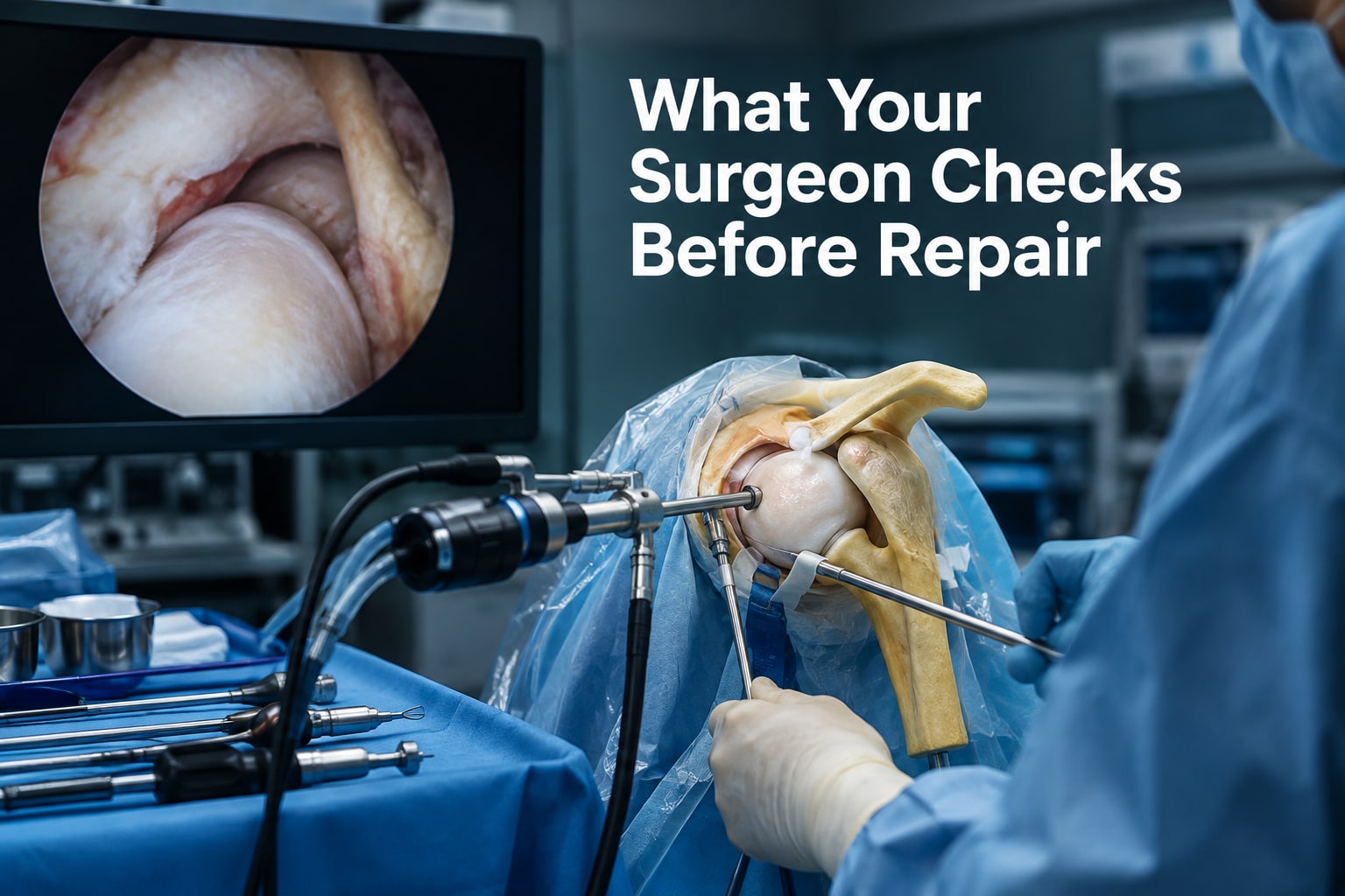
After anesthesia, the surgeon sets the arm in a chosen position, usually beach-chair or lateral, so the shoulder can be viewed and moved safely. Small incisions create the entry points.
Through a posterior portal, the surgeon inserts the arthroscope camera and fills the joint with sterile saline irrigation fluid, which expands the working space and clears blood and debris.
The initial survey follows a planned sequence rather than jumping to the abnormality named on an MRI report:
- In the glenohumeral joint, the surgeon rotates the camera to inspect the joint surfaces, labrum, biceps attachment, capsule, synovium and visible rotator cuff from inside.
- The surgeon uses an anterior portal and accessory portals in shoulder arthroscopy for instruments, lighting angles and probing. A probe tests whether the labrum and other attachments are firmly fixed or pull away abnormally.
- After the joint survey, the surgeon withdraws and redirects the camera into the subacromial space. The bursa can obscure the view, so removing inflamed bursal tissue may be necessary before checking the outer rotator cuff and acromion.
- The surgeon moves the arm while watching the tissues. This reveals contact, tendon movement, labral instability or capsular looseness that a still image cannot show.
This two-compartment inspection reduces the risk of treating one visible lesion while missing a second problem elsewhere.
What the surgeon checks inside the glenohumeral joint
Inside the glenohumeral joint, the surgeon inspects cartilage, the labrum, biceps attachment, cuff undersurface, capsule and synovium—not just the tear suspected on an MRI.
| Structure | What is inspected | Abnormalities identified |
|---|---|---|
| Humeral head and glenoid | Humeral head cartilage and glenoid cartilage for surface continuity | Wear, fissures, flaps, exposed bone and loose bodies; this damage can reduce the benefit of treating only a tendon or labrum |
| Labrum and biceps | The labrum around its full circumference, including the biceps anchor and long-head biceps tendon | An anterior-inferior Bankart tear, a superior SLAP tear, posterior labral damage, fraying or an unstable attachment |
| Rotator cuff articular surface | The joint-facing surfaces of the supraspinatus, infraspinatus and subscapularis | Partial-thickness tears, with their depth, location and stability assessed; the upper and lower subscapularis borders and biceps pulley receive particular attention |
| Capsule and synovium | The joint lining and capsule for excess tissue, inflammation and abnormal tightness | Synovitis, scar tissue, capsular thickening or restrictions consistent with adhesive capsulitis |
A probe helps distinguish a stable anatomical labral variant from a detached or unstable tear. A small, stable frayed cuff area is not automatically equivalent to a deep partial tear requiring repair.
Multiple findings can coexist, such as cuff damage with biceps disease, labral injury or cartilage wear, so the operative choice depends on which abnormalities match your symptoms and examination.
What is examined in the subacromial space and during arm movement
After entering the subacromial space, the surgeon clears inflamed tissue when it blocks the view and examines the structures that can cause pain or restrict movement:
- The subacromial bursa is checked for thickening, bleeding, or bursitis. Removing part of it, called a bursectomy, improves visibility but does not itself treat every pain source.
- The outer rotator cuff is inspected for fraying or a bursal-sided rotator cuff tear. A partial-thickness rotator cuff tear is judged by its depth, location, and stability; a small unstable flap is not automatically a full-thickness tear requiring repair.
- The acromion undersurface is assessed for a spur, rough edge, or narrowing that could contribute to impingement. Acromioplasty is not automatic; the surgeon performs it only when the shape and mechanical contact support that decision.
Arm movement adds information a static scan cannot provide. During this dynamic assessment, the surgeon rotates or elevates the arm while watching whether the cuff rubs against the acromion, whether a tendon catches, and whether the tissues remain stable under load.
Probing can also expose capsular laxity or excessive translation that is absent while the shoulder lies still.
Movement shows restriction as well. A shoulder that cannot rotate or elevate despite no large tear may fit adhesive capsulitis, while abnormal looseness points toward instability. The surgeon also checks the subscapularis at its upper and lower borders near the biceps pulley, where a subtle tear can hide.
How arthroscopy findings are matched with MRI and symptoms
MRI versus arthroscopy can produce different answers because MRI shows structures in fixed positions, while the camera reveals their actual surface, stability and tissue quality. Arthroscopy may confirm a tear, expose cartilage damage or synovitis, or show that an apparent defect is less significant than the scan suggested.
The surgeon does not treat every visible abnormality. Age-related cartilage wear and incidental labral fraying are common findings that may not explain pain, weakness or restricted shoulder function. A defect matters when its location and severity fit your history, physical examination, movement limits and the mechanism of injury.
For suspected instability, the surgeon matches the appearance to the direction and pattern of giving-way episodes:
- An anterior-inferior Bankart tear fits repeated anterior dislocation or slipping.
- A posterior labral tear fits posterior instability and pain with pushing or loading.
- A HAGL lesion supports a humeral-sided capsular avulsion after instability.
- Capsular laxity supports looseness; adhesive capsulitis and capsular contracture instead explain global stiffness and loss of motion.
Arthroscopy also helps identify combined problems, such as a cuff tear with biceps disease or cartilage damage. Major glenoid bone loss remains a measurement problem: a CT scan, along with MRI, may be needed to plan instability treatment because the camera cannot define the entire bony shape reliably.
The final diagnosis therefore reflects the scan, direct inspection and how closely each finding reproduces your symptoms.
How the inspection leads to repair, debridement or no treatment
The surgeon chooses repair when a lesion is unstable, structurally important and consistent with your symptoms; frayed tissue without mechanical significance is more often treated with debridement or left alone. The decision also follows tissue quality, tear size, instability and the strength of available fixation.
| Option | What it addresses | When it applies |
|---|---|---|
| Labral repair | Reattaches a detached labrum | Traumatic instability or a symptomatic tear with sound tissue |
| Rotator cuff repair | Restores a torn tendon | A repairable tear that matches weakness and pain |
| Debridement | Removes loose or frayed tissue | Stable partial tears, ragged labrum or inflamed synovium |
| Bursectomy | Clears inflamed bursal tissue | When the bursa blocks a clear view or causes persistent impingement |
| Acromioplasty | Reshapes the acromion | Selected narrowing or spur-related contact, not automatically |
| Capsular release | Divides a tight capsule | Adhesive capsulitis limiting motion |
The biceps anchor is treated separately from the labrum when the long-head tendon is frayed, partly torn, displaced or affected by pulley instability. Biceps tenodesis moves and fixes the tendon outside the joint; biceps tenotomy releases it without fixation. Either may be more suitable than labral repair alone.
Before adding procedures, the surgeon checks the surgical consent and confirms that the finding justifies the extra treatment. Dr Aniruddha Deshmukh-Orthopedic Doctor, Joint Replacement & Arthroscopy Surgeon in Hinjewadi, Pune, can use this correlation to avoid treating an incidental abnormality.
The final arthroscopic check confirms stable fixation, secure suture anchors, smooth motion and no hardware prominence, inadequate fixation or excessive tightening. The surgeon also checks the repaired cuff, labrum, biceps treatment and capsule before closing.
Related services
Arthroscopy Surgeon Arthroscopy Surgeon in Hinjewadi, Pune Dr. Aniruddha Deshmukh – Leading Arthroscopy Surgeon in Hinjewadi, Marunji and Pune. Dr. |
Arthroscopy Surgeon Arthroscopy Surgeon in Mahalunge, Pune Dr. Aniruddha Deshmukh – Leading Arthroscopy Surgeon in Mahalunge, Sus, Pashan and Pune. Dr. |
Frequently asked questions
How does a surgeon enter and survey the shoulder during arthroscopy?
After anesthesia, the surgeon positions the arm in a beach-chair or lateral position and uses small incisions for the camera and instruments. The camera provides a systematic view of the joint before treatment begins.
What does the surgeon check inside the glenohumeral joint?
The inspection includes the articular cartilage, labrum, rotator cuff undersurface, biceps tendon and anchor, joint capsule, and signs of instability or inflammation.
What is examined in the subacromial space and during arm movement?
The surgeon checks the bursa, acromion, bone spurs, coracoacromial ligament and outer surface of the rotator cuff. Moving the arm helps assess impingement, tendon contact and instability.
How are arthroscopy findings matched with MRI and symptoms?
The surgeon compares what the camera shows with the MRI location and the symptoms you reported. A tear on MRI is treated only when its appearance and position fit the clinical findings.
How do inspection findings determine repair, debridement or no treatment?
Repair is chosen when tissue damage needs structural restoration. Debridement removes frayed or inflamed tissue when repair is unnecessary. Stable, minor or unrelated findings can require no treatment.
Related services
Arthroscopy Surgeon Arthroscopy Surgeon in Pimple Saudagar, Pune Dr. Aniruddha Deshmukh – Leading Arthroscopy Surgeon in Pimple Saudagar, Pimple Gurav, Rahatani and Pune. Dr. |
Arthroscopy Surgeon Arthroscopy Surgeon in Wakad, Pune Dr. Aniruddha Deshmukh – Leading Arthroscopy Surgeon in Wakad, Pimple Nilakh, Thergaon and Pune. Dr. |
Arthroscopy Surgeon Arthroscopy Surgeon in Baner, Balewadi, Pune Dr. Aniruddha Deshmukh – Leading Arthroscopy Surgeon in Baner, Balewadi, and Pune. Dr. View service → |
Arthroscopy Surgeon Arthroscopy Surgeon in Punawale, Pune Dr. Aniruddha Deshmukh – Leading Arthroscopy Surgeon in Punawale and Pune. Dr. |