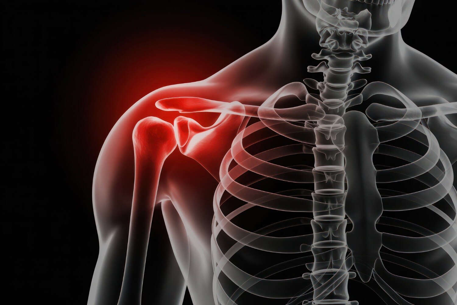
A fall, road accident, sports injury or direct blow can damage several structures around the shoulder, and pain alone cannot identify which one is injured. You will learn how urgent danger is recognised, how the arm is examined and imaged, and how a surgeon chooses between immobilisation, rehabilitation, fixation or replacement.
Key takeaways
- Seek emergency care for an open fracture, severe deformity, numbness, or a cold hand.
- Tell the surgeon exactly how the injury happened and when symptoms began.
- X-rays show bone alignment; CT maps complex fractures, while MRI assesses soft tissues.
- Follow-up checks healing, shoulder movement, strength, and readiness for daily activities.
What happens during the first shoulder trauma assessment?
The first assessment reconstructs how the injury happened: a fall onto an outstretched hand, direct blow, road accident or forceful pull creates different patterns. The surgeon asks when the pain and deformity began, then examines the patient before relying on pain location or an X-ray.
The examination works through these checks:
- The skin is checked for an open wound, bruising, swelling or bone threatening to break through. The shoulder contour is compared with the uninjured side for flattening, prominence or abnormal position.
- Active and passive movement is tested only where safe. Forced movement is avoided when a fracture or dislocation is obvious.
- The hand is checked for temperature, colour, capillary refill and distal wrist pulses. A normal pulse confirms blood is reaching the hand, but it does not exclude nerve damage.
- Sensation over the lateral shoulder is tested for possible axillary nerve injury. Radial nerve function includes wrist and finger extension; ulnar nerve function includes finger spreading and closing.
- The surgeon separates a shoulder fracture from a dislocation, acromioclavicular joint injury, collarbone fracture, upper-arm fracture and soft-tissue injury. Radiographs, and sometimes CT or MRI, confirm what examination alone cannot.
Pain over the shoulder does not establish the diagnosis; neck pain can refer there, and tendon injury can produce severe pain without a broken bone. This structured assessment guides safe shoulder fracture treatment in Pune.
Which shoulder injuries need emergency assessment?
Emergency assessment is needed for any shoulder injury with an open fracture, uncontrolled bleeding, visible deformity, skin tenting, an absent wrist pulse, a pale or blue hand, cold fingers, rapidly increasing swelling, new numbness or weakness, or severe loss of function.
Do not wait for a routine appointment; these signs can indicate damage to bone, skin, nerves, or blood vessels.
- Cover an open wound with a clean dressing without pushing exposed bone back inside.
- Keep the arm supported in the position found.
- Remove rings, watches, and tight clothing before swelling increases.
- Do not force a deformed shoulder back into place.
- Do not immobilise it in a new position without medical assessment.
A dislocated shoulder generally needs prompt reduction by a trained clinician. The clinician should record sensation over the lateral shoulder and check distal pulses before and after reduction, because an axillary-nerve or vascular injury can accompany the dislocation.
| Finding | Why it matters | Correct response |
|---|---|---|
| Open fracture or uncontrolled bleeding | Infection, blood loss, and tissue damage risk | Emergency department assessment |
| Deformity or skin tenting | Bone may be displaced against skin or surrounding tissue | Do not manipulate; seek emergency care |
| Absent pulse, pale or blue hand, or cold fingers | Possible arterial compromise | Immediate emergency assessment |
| New numbness or weakness | Possible nerve injury, even with a normal pulse | Urgent examination |
| Severe loss of function or rapidly increasing swelling | Possible fracture-dislocation or concealed bleeding | Emergency assessment |
If you search “shoulder surgeon pimple saudagar,” use the same red-flag standard: locality does not change the urgency. A shoulder surgeon must assess the injury before reduction or immobilisation so an associated fracture, nerve injury, or blood-vessel injury is not missed or worsened.
How do X-rays, CT and MRI reveal the actual injury?
Plain shoulder radiographs are usually the first test because they are quick and show bone alignment, fracture, and dislocation. An anteroposterior view alone is insufficient: overlapping structures can hide displacement or a humeral-head dislocation. Ask for axillary and scapular-Y views, which show the shoulder from different angles and clarify joint position.
| Test | What it reveals | Main value |
|---|---|---|
| X-ray | Proximal humerus or clavicle fracture, acromioclavicular separation, dislocation | Fast initial assessment of alignment and bone injury |
| CT | Complex fracture lines, joint-surface involvement, bone fragments and three-dimensional anatomy | Defines operative planning when a fracture is displaced or comminuted |
| MRI | Rotator-cuff tears, labral injury, muscle damage and occult bone injury | Explains persistent symptoms when the X-ray does not show the cause |
A proximal humerus fracture may be minimally displaced, while a clavicle fracture may shorten or shift; an acromioclavicular separation changes the joint contour, and a dislocation moves the humeral head out of its socket. CT maps these patterns rather than simply confirming that a fracture exists.
MRI is useful when pain, weakness or restricted movement persists despite reassuring radiographs.
If you search for fracture trauma care balewadi in baner, ask whether imaging includes the necessary views and whether CT or MRI is being recommended for a specific unanswered question. Pain location alone cannot distinguish a fracture from tendon injury or referred neck pain.
How is the choice between a sling and surgery made?
An X-ray showing a fracture does not automatically mean surgery. Many minimally displaced proximal humerus and clavicle fractures heal in a sling, shoulder immobiliser or collar-and-cuff, followed by supervised motion to limit stiffness.
Surgery becomes more likely when the pattern is displaced, unstable, involves the joint surface, is open, or includes a fracture-dislocation. The decision depends on:
- Angulation, shortening and loss of alignment
- Instability or a fracture-dislocation
- Bone quality and the likelihood of holding a repair
- Age and activity demands
- Medical fitness for anaesthesia and surgery
- Nerve or blood-vessel damage
- Threatened skin, particularly over a displaced clavicle
A surgeon may use plate-and-screw fixation for a clavicle or proximal humerus fracture, or intramedullary nailing for selected upper-arm patterns. Selected older adults need shoulder replacement when the humeral head cannot be reliably reconstructed; reverse shoulder arthroplasty is an option when the rotator cuff is insufficient, not a default treatment for every fracture.
Non-operative care avoids anaesthetic complications, infection and prominent hardware. Fixation can restore alignment but may leave visible or irritating metal, with later implant removal sometimes required. Prolonged immobilisation creates its own problem: shoulder stiffness that becomes difficult to reverse.
Searches for shoulder surgeon fracture trauma care pimple saudagar or fracture trauma care mahalunge describe location, not different treatment standards. The same alignment, nerve, circulation and functional assessment guides the choice.
What does complete recovery and follow-up involve?
Recovery continues after the shoulder is reduced or repaired surgically. Follow-up X-rays check whether the bones remain aligned, whether the fracture has lost position, and whether healing is progressing.
| Follow-up finding | Rehabilitation response | Why it matters |
|---|---|---|
| Stable alignment and healing | Increase supervised movement gradually | The repair can tolerate more activity |
| Loss of position or delayed healing | Restrict loading and reassess treatment | Early lifting can jeopardise union |
| Stiffness without instability | Add guided mobility work | Excessive immobilisation can make stiffness difficult to reverse |
Your programme must match the stability of the fracture fixation or repair. Do not lift actively, carry weight, or push up from a chair until the treating surgeon permits it; a shoulder that feels better can still have an unhealed fracture.
When comparing shoulder fracture treatment in Pune, look for a complete local pathway that can coordinate:
- Emergency assessment and appropriate X-rays or CT
- Reduction when a fracture-dislocation requires it
- Fixation or shoulder replacement when the injury pattern justifies that choice
- Documented circulation, sensation and motor checks before and after treatment
- Structured rehabilitation with scheduled review
Dr Aniruddha Deshmukh-Orthopedic Doctor, Joint Replacement & Arthroscopy Surgeon in Hinjewadi, Pune is one local option to assess whether these stages are coordinated rather than treating surgery as the entire plan. Many injuries heal without surgery; others need fixation or replacement because of displacement, instability, joint-surface damage or an unreconstructible humeral head.
Related services
Fracture and Trauma Care Fracture and Trauma Care Specialist in Hinjewadi, Pune Dr. Aniruddha Deshmukh – Trusted Fracture and Trauma Care Specialist in Hinjewadi, Marunji and Pune. View service → |
Fracture and Trauma Care Fracture and Trauma Care Specialist in Pimple Saudagar, Pune Dr. Aniruddha Deshmukh – Trusted Fracture and Trauma Care Specialist in Pimple Saudagar, Pimple Gurav, Rahatani and Pune. |
Frequently asked questions
What happens during the first shoulder trauma assessment?
The surgeon asks how the injury happened, when pain or deformity began, and examines the shoulder, arm, skin, circulation, sensation, and movement before ordering imaging.
Which shoulder injuries need emergency assessment?
Seek urgent assessment for an open wound over a fracture, obvious deformity, uncontrolled bleeding, severe swelling, numbness, weakness, loss of circulation, or a hand that feels cold or looks pale or blue.
How do X-rays, CT and MRI reveal the actual injury?
X-rays show fractures, dislocations, and alignment. CT creates detailed cross-sectional views for complex fracture patterns. MRI evaluates tendons, ligaments, cartilage, muscles, and some hidden fractures.
How is the choice between a sling and surgery made?
The decision depends on fracture location, displacement, joint involvement, shoulder stability, nerve or blood-vessel injury, activity demands, medical risks, and whether the bones can heal in a useful position.
What does complete recovery and follow-up involve?
Follow-up tracks bone healing, pain, shoulder range of motion, strength, and function. Your plan may progress from protection to supervised exercises and task-specific rehabilitation.
Related services
Fracture and Trauma Care Fracture and Trauma Care Specialist in Wakad, Pune Dr. Aniruddha Deshmukh – Trusted Fracture and Trauma Care Specialist in Wakad, Pimple Nilakh, Thergaon and Pune. View service → |
Shoulder and Elbow Pain Treatment Shoulder and Elbow Pain Treatment in Wakad, Pune Dr. Aniruddha Deshmukh is a trusted Fracture and Trauma Care Specialist in Wakad, Pimple Nilakh, Thergaon and Pune, providing comprehensive... View service → |
Fracture and Trauma Care Fracture and Trauma Care Specialist in Mahalunge, Pune Dr. Aniruddha Deshmukh – Trusted Fracture and Trauma Care Specialist in Mahalunge, Sus, Pashan and Pune. View service → |
Fracture and Trauma Care Fracture and Trauma Care Specialist in Baner, Balewadi, Pune Dr. Aniruddha Deshmukh – Trusted Fracture and Trauma Care Specialist in Baner, Balewadi and Pune. View service → |